
MED ED ONLINE
Apprendre à bien manger en ligne
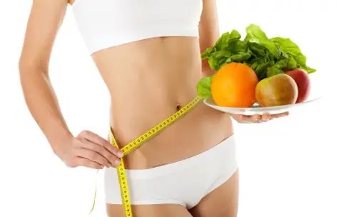
Coaching en nutrition
Med Ed Online vous propose des coachings nutritionnels pour retrouver votre équilibre alimentaire
Résultats garantis
En suivant nos conseils, nous vous garantissons que vous perdrez du poids ou retrouverez un équilibre alimentaire sain.
Accompagnement personnalisé
Si vous le souhaitez, nous pouvons vous accompagner personnellement pour atteindre vos objectifs.
Apprenez à manger seinement sans changer votre style de vie
Manger sainement n’a pas la même définition pour tous. Pour certains cela va être de manger ni trop gras ni trop salé, pour certains de manger sans gluten et pour d’autres de manger vegan. Parce que pour chaque individu il existe une façon différente de manger sainement, Med Ed Online aborde tous les sujets liés à la nutrition, pour que chacun y trouve son compte.
Vous avez du mal à trouver la bonne balance nutritionelle ?
Vous n’êtes pas tout seul ! Nous sommes là pour vous aider.
La science de la nutrition est très jeune. Entre le différents loby agroalimentaires et les fausses informations et croyances qui circulent sur internet, il n’est clairement pas facile de s’y retrouver. Si à cela on ajoute les différentes études scientifiques qui vont jusqu’à se contredire, on peut rapidement être complètement perdu.
C’est pourquoi l’équipe de Med Ed Online, vous accompagne en démêlant le vrai du faux et vous aide à trouver la réponse à toutes vos questions liées à la nutrition
Comment nous pouvons vous aider ?
L’équipe Med Ed Online vous propose quotidiennement des nouveaux articles pour vous aider à mieux manger et à vous sentir mieux dans votre corps. Voici les principaux thèmes que nous abordons.
Coaching nutritionnel
Comment changer ses habitudes alimentaires et mieux manger ?
Nutrition sportive
Comment bien s’alimenter lorsque l’on fait du sport ?
Vitamines et compléments
Quelles vitamines ou compléments alimentaires
Perte de poids
Comment perdre du poids de manière efficace et durable ?


Trouver une balance peut être difficile mais manger sainement n’a pas a l’être
Avec nos vies à 100 à l’heure, il est parfois difficile de trouver le temps de bien manger. On ne prend plus forcément le temps de faire les courses et de prendre une longue pause déjeuner le midi. Mais même les personnes disposant de peu de temps peuvent se nourir correctement.
A propos d’Emma
Coach en Nutrition
Je m’appelle Emma et je suis coach en nutrition. Depuis une dizaine d’années environ, j’accompagne mes clients dans leur quête d’une alimentation plus saine et équilibrée. Il y a peu, j’ai décidé de lancer Med-Ed-Online, afin de rendre accessible au plus grand nombre mes conseils sur la nutrition et la perte de poids, principale source d’inquiétude de mes clients. Bonne lecture !

Les coupe faim

Les bruleurs de graisses

Il existe des centaines de bruleurs de graisses et il n’est pas facile de s’y retrouver. Voici les meilleurs brule graisse
Les régimes

Comment faire un régime et ne pas reprendre du poids ? On vous explique tout avec les régimes efficaces
Nous avons développé une méthode révolutionnaire pour bien manger
Après des années de travail, nous sommes fiers de vous présenter notre méthode qui vous permet de changer facilement vos habitudes alimentaires et d’en finir avec la junk food.
Vous apprendrez enfin à bien manger et à rééquilibrer votre alimentation.
Autres ressources gratuites
Calculez votre IMC aujourd’hui
Car pour calculer son IMC il ne suffit pas de donnerson poids et sa taille, contacter nous pour un IMC et des recommandations personnalisées.
« J’ai découvert Med Ed Online il y a peu et depuis que je consulte le site régulièrement je dois avouer que je mange beaucoup mieux et que ma forme quotidienne est au beau fixe. J’ai perdu 2 kilos. »
« Toujours pressé, jamais le temps de me faire à manger ou bien de faire les courses. Sur Med Ed Online j’ai trouvé plein d’astuces pour bien manger quand on est pressé et j’avoue que ça fait du bien. »
Les derniers articles du blog
Pour manger mieux et perdre du poids

Le CBD : quel apport dans la perte de poids ?
Le cannabidiol, ou CBD, est une molécule non psychotrope et non addictive extraite de la plante Cannabis L. Sativa, également appelée chanvre cultivé. Cette molécule librement commercialisée dans l’Hexagone pourrait contribuer à la perte de poids. CBD : les raisons...

Les bienfaits de la vitamine D sur l’organisme
La vitamine D, également appelée vitamine du soleil, est une substance indispensable pour notre organisme, car elle assure notre bonne santé physique et morale. Pourtant, nous la négligeons souvent, à tel point que nous finissons par souffrir d’une carence. Dans le...
Meilleur brûleur de graisse puissant et efficace : mon TOP 7
Perdre du poids est pour certaines personnes un vrai défi ! Pour atteindre son objectif minceur, la détermination, la motivation et la volonté sont les maîtres mots. Mais aujourd'hui, pour vous aider à maigrir vite et plus facilement, des brûleurs de graisses sont...